Most ebook files are in PDF format, so you can easily read them using various software such as Foxit Reader or directly on the Google Chrome browser.
Some ebook files are released by publishers in other formats such as .awz, .mobi, .epub, .fb2, etc. You may need to install specific software to read these formats on mobile/PC, such as Calibre.
Please read the tutorial at this link. https://ebooknice.com/page/post?id=faq
We offer FREE conversion to the popular formats you request; however, this may take some time. Therefore, right after payment, please email us, and we will try to provide the service as quickly as possible.
For some exceptional file formats or broken links (if any), please refrain from opening any disputes. Instead, email us first, and we will try to assist within a maximum of 6 hours.
EbookNice Team
Status:
Available0.0
0 reviews
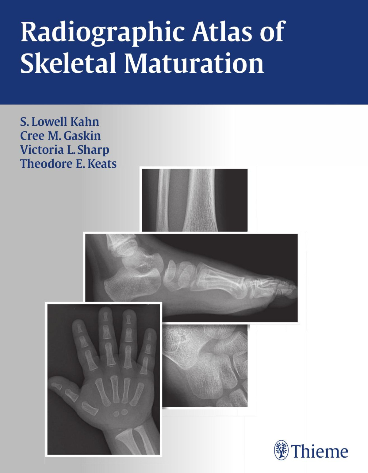
ISBN 10: 1604065710
ISBN 13: 9781604065718
Author: S Lowell Kahn, Christopher M Gaskin, Victoria L Sharp, Theodore E Keats, Bing Li
Nearly 2,300 images provide the reference standard for normal skeletal maturation at every developmental stage
When dealing with the maturing skeleton and its many complex growth alterations, physicians are constantly faced with the question: "Is this image normal?" The Radiographic Atlas of Skeletal Maturation succinctly answers that question by providing a comprehensive set of male and female reference images for every age and body part. This allows physicians to quickly hone in on "normal" ranges for the specific case they are reviewing--particularly useful when called upon to read a pediatric skeletal radiograph in the emergency room or while on call.
Special Features:
Access to nearly 2,300 high-quality images that provide instant reference to "normal" views of the skeleton at every developmental milestone-available in both the text and accompanying DVD
Multiple projections at every age, sex, and body part combination so that the user can match the reference points in the book to the case at hand and arrive at a solid clinical interpretation
Practical text layout organized by gender and body part that provides quick access to images of normal development at any given age
A software virtual "skeletal survey" demonstrates images of younger and older individuals and crystallizes the subtle variations in growth patterns
Powerful software package with advanced image enhancement tools allows optimization of atlas image details for greater clarity. Compatible with numerous image formats (including DICOM) allowing viewing and editing of outside images
Convenient growth charts included in the book and DVD
This unique resource, with its vast collection of print and DVD images of normal progressive skeletal development, gives physicians the full range of comparative information they need to interpret pediatric skeletal radiographs in any clinical setting. It is the reference standard for radiologists, pediatricians, orthopedists, emergency room physicians, internists, rehabilitation physicians, and training physicians who are called upon to review a pediatric radiograph and confidently make a diagnosis.
Section I. Male
Male Skull
Male Cervical Spine
Male Thoracic Spine
Male Lumbar Spine
Male Chest
Male Shoulder
Male Humerus
Male Elbow
Male Forearm
Male Wrist
Male Hand
Male Pelvis
Male Femur
Male Knee
Male Tibia and Fibula
Male Ankle
Male Foot
Section II. Female
Female Skull
Female Cervical Spine
Female Thoracic Spine
Female Lumbar Spine
Female Chest
Female Shoulder
Female Humerus
Female Elbow
Female Forearm
Female Wrist
Female Hand
Female Pelvis
Female Femur
Female Knee
Female Tibia and Fibula
Female Ankle
Female Foot
Appendix
radiographic atlas of skeletal maturation
radiographic atlas of skeletal maturation pdf
radiographic atlas of skeletal development of the hand and wrist
radiographic atlas of skeletal development
radiographic atlas of skeletal development of the knee
radiographic evaluation of skeletal maturation
Tags: S Lowell Kahn, Christopher M Gaskin, Victoria L Sharp, Theodore E Keats, Bing Li, Radiographic, Atlas

4.6
12 reviews

4.7
9 reviews

4.8
7 reviews
4.5
20 reviews
4.5
12 reviews