logo
Product categories

EbookNice.com

Most ebook files are in PDF format, so you can easily read them using various software such as Foxit Reader or directly on the Google Chrome browser.
Some ebook files are released by publishers in other formats such as .awz, .mobi, .epub, .fb2, etc. You may need to install specific software to read these formats on mobile/PC, such as Calibre.

Please read the tutorial at this link.  https://ebooknice.com/page/post?id=faq


We offer FREE conversion to the popular formats you request; however, this may take some time. Therefore, right after payment, please email us, and we will try to provide the service as quickly as possible.


For some exceptional file formats or broken links (if any), please refrain from opening any disputes. Instead, email us first, and we will try to assist within a maximum of 6 hours.

EbookNice Team
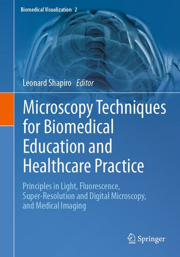
(Ebook) Microscopy Techniques for Biomedical Education and Healthcare Practice: Principles in Light, Fluorescence, Super-Resolution and Digital Microscopy, and Medical Imaging by Leonard Shapiro, (ed.) ISBN 9783031368493, 3031368495

  • SKU: EBN-52617030
Zoomable Image
$ 32 $ 40 (-20%)

Status:

Available

0.0

0 reviews
Instant download (eBook) Microscopy Techniques for Biomedical Education and Healthcare Practice: Principles in Light, Fluorescence, Super-Resolution and Digital Microscopy, and Medical Imaging after payment.
Authors:Leonard Shapiro, (ed.)
Pages:242 pages.
Year:2023
Editon:1st ed. 2023
Publisher:Springer
Language:english
File Size:89.56 MB
Format:epub
ISBNS:9783031368493, 3031368495
Categories: Ebooks

Product desciption

(Ebook) Microscopy Techniques for Biomedical Education and Healthcare Practice: Principles in Light, Fluorescence, Super-Resolution and Digital Microscopy, and Medical Imaging by Leonard Shapiro, (ed.) ISBN 9783031368493, 3031368495

This edited book has a strong focus on advances in microscopy that straddles research, medical education and clinical practice. These advances include the shift in power from conventional to digital microscopy. The first section of this book covers imaging techniques and morphometric image analysis with its applications in biomedicine using different microscopy modes. Chapters highlight the rich development of fluorescence methods and technologies; particle tracking techniques with applications in biomedical research and nanomedicine; the way in which visualizations have revolutionized taxonomy from gross anatomy to genetics; and the psychology of perception and how it affects our understanding of cells and tissues. The book’s first section concludes by exploring the use of CT modalities to evaluate anterior deformities in craniosynostosis. In the second section of the book, chapters on anatomical and cell biology education explore the history of anatomical models and their use in educational settings. This includes examples in 3D printing and functional human anatomical models that can be created using easily available resources and the use of biomedical imaging in visuospatial teaching of anatomy; the novel use of ultrasound in medical education and practice; and skill acquisition in histology education using a flowchart called a ‘decision tree’. This book will appeal to histologists, microscopists, cell biologists, clinicians and those involved in anatomical education and biomedical visualization, as well as students in those respective fields.
*Free conversion of into popular formats such as PDF, DOCX, DOC, AZW, EPUB, and MOBI after payment.

Related Products