logo
Product categories

EbookNice.com

Most ebook files are in PDF format, so you can easily read them using various software such as Foxit Reader or directly on the Google Chrome browser.
Some ebook files are released by publishers in other formats such as .awz, .mobi, .epub, .fb2, etc. You may need to install specific software to read these formats on mobile/PC, such as Calibre.

Please read the tutorial at this link.  https://ebooknice.com/page/post?id=faq


We offer FREE conversion to the popular formats you request; however, this may take some time. Therefore, right after payment, please email us, and we will try to provide the service as quickly as possible.


For some exceptional file formats or broken links (if any), please refrain from opening any disputes. Instead, email us first, and we will try to assist within a maximum of 6 hours.

EbookNice Team
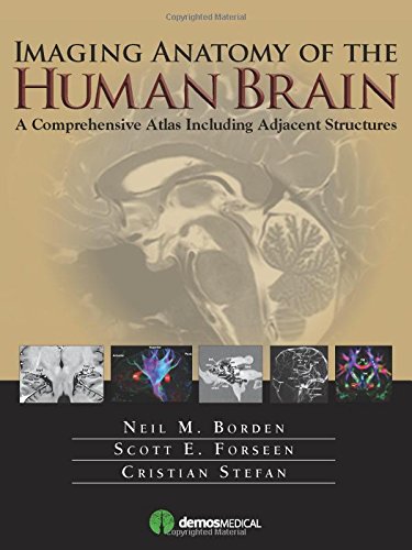
(Ebook) Imaging Anatomy of the Human Brain: A Comprehensive Atlas Including Adjacent Structures by Neil M. Borden, Scott E. Forseen, Cristian Stefan ISBN 9781936287741, 1936287749

  • SKU: EBN-5252372
Zoomable Image
$ 32 $ 40 (-20%)

Status:

Available

4.5

22 reviews
Instant download (eBook) Imaging Anatomy of the Human Brain: A Comprehensive Atlas Including Adjacent Structures after payment.
Authors:Neil M. Borden, Scott E. Forseen, Cristian Stefan
Pages:464 pages.
Year:2015
Editon:1
Publisher:Demos Medical
Language:english
File Size:50.47 MB
Format:pdf
ISBNS:9781936287741, 1936287749
Categories: Ebooks

Product desciption

(Ebook) Imaging Anatomy of the Human Brain: A Comprehensive Atlas Including Adjacent Structures by Neil M. Borden, Scott E. Forseen, Cristian Stefan ISBN 9781936287741, 1936287749

The most precise, cutting-edge images of normal cerebral anatomy available today are the centerpiece of this spectacular atlasfor clinicians, trainees, and students in the neurologically-based medical and non-medical specialties. Truly an ìatlas for the 21st century,î this comprehensive visual reference presents a detailed overview of cerebral anatomy acquired through the use of multiple imaging modalities including advanced techniques that allow visualization of structures not possible with conventional MRI or CT. Beautiful color illustrations using 3-D modeling techniques based upon 3D MR volume data sets further enhances understanding of cerebral anatomy and spatial relationships. The anatomy in these color illustrations mirror the black and white anatomic MR images presented in this atlas. Written by two neuroradiologists and an anatomist who are also prominent educators, along with more than a dozen contributors, this state-of-the-art atlas will serve as an authoritative learning tool in the classroom, and as an invaluable practical resource at the workstation or in the office or clinic.
*Free conversion of into popular formats such as PDF, DOCX, DOC, AZW, EPUB, and MOBI after payment.

Related Products