Most ebook files are in PDF format, so you can easily read them using various software such as Foxit Reader or directly on the Google Chrome browser.
Some ebook files are released by publishers in other formats such as .awz, .mobi, .epub, .fb2, etc. You may need to install specific software to read these formats on mobile/PC, such as Calibre.
Please read the tutorial at this link. https://ebooknice.com/page/post?id=faq
We offer FREE conversion to the popular formats you request; however, this may take some time. Therefore, right after payment, please email us, and we will try to provide the service as quickly as possible.
For some exceptional file formats or broken links (if any), please refrain from opening any disputes. Instead, email us first, and we will try to assist within a maximum of 6 hours.
EbookNice Team
Status:
Available4.4
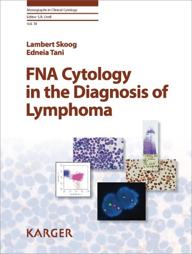
40 reviewsISBN 10: 3805586264
ISBN 13: 978-3805586269
Author: Lambert Skoog, Edneia Tani, Anja Porwitt
Fine-needle aspiration (FNA) became important for the cytology of the enlarged lymph node in the 1950s and 1960s and was accepted in the diagnosis of various types of lymphadenitis and metastatic disease. The diagnosis of lymphoma by FNA cytology, however, remained controversial for many years, as FNA smears did not allow the evaluation of growth pattern. Only later with the introduction of immunocytochemistry on FNA material it became possible to conclusively diagnose the majority of lymphomas with an accuracy comparable to that of histopathology. Other ancillary techniques such as FISH and PCR can now also be applied successfully to FNA material. These facts together with the excellent clinical performance of FNA sampling should increase the spread of the technique. This comprehensive manual presents the cytomorphologic and immunocytochemical characteristics of both non-Hodgkin and Hodgkin lymphomas. It discusses the technical, methodological aspects of lymphoma diagnosis and describes the cytologic features of reactive lymphoid lesions and the major types of neoplastic lymphoid lesions, based on the most recent (2001) WHO lymphoma classification. Key cytologic and immunologic features are listed to facilitate a conclusive diagnosis of the different lesions. This publication will be of immense value to clinicians such as cytopathologists, pathologists, oncologists, and hematologists involved in the clinical work-up and management of patients with lymph node lesions.
Chapter 1
Historical Aspects
General Aspects
References
Chapter 2
Techniques
Fine Needle Aspiration Biopsy and Smear Preparation
Fixation and Staining
Fluid Preparation
Cytospin Preparation
Storage
Immunostaining
Cell Proliferation
Molecular Techniques
References
Chapter 3
Flow Cytometry in Fine-Needle Aspiration Diagnosis of Lymphomas In collaboration with Anja Porwit
Methodological Considerations
Sample Preparation
Antibody Panel
Data Analysis
Advantages and Disadvantages of FC
Comparison between FC and Immunocytochemistry on Cytospins
How to Get the Best Results in FC Diagnostics of FNA
References
Chapter 4
B Cell Neoplasms
WHO Histological Classification of B Cell Neoplasms
Mature B Cell Neoplasms
Precursor B Cell Neoplasm
Small Lymphocytic Lymphoma/Chronic Lymphocytic Leukemia
B Cell Prolymphocytic Leukemia
Lymphoplasmacytic Lymphoma
Splenic Marginal Zone Lymphoma
Hairy Cell Leukemia
Plasma Cell Neoplasms
(1) Myeloma
(2) Plasmacytoma (Osseous/Extraosseous)
Marginal Zone Lymphoma/Extranodal (MALT) and Nodal
Follicular Lymphomas
Mantle Cell Lymphoma
Diffuse Large B Cell Lymphoma
Mediastinal Large B Cell Lymphoma
Burkitt Lymphoma
Lymphomatoid Granulomatosis
Precursor B Lymphoblastic Leukemia/Lymphoma
References
Chapter 5
T Cell Neoplasms
WHO Histological Classification of Mature T Cell and NK Cell Neoplasms
T Prolymphocytic Leukemia
Adult T Cell Lymphoma/Leukemia
Mycosis Fungoides and Sezary Syndrome
Extranodal NK/T Cell Lymphoma, Nasal Type
Angioimmunoblastic T Cell Lymphoma
Peripheral T Cell Lymphoma, Unspecified
Anaplastic Large Cell (CD30+) Lymphoma
Precursor T Cell Leukemia/Lymphoma
References
Chapter 6
Hodgkin Lymphoma
WHO Histological Classification, Hodgkin Lymphoma
Classical Hodgkin Lymphoma
(1) Nodular Sclerosis Variant
(2) Mixed Cellularity Variant
(3) Lymphocyte-Rich Variant
(4) Lymphocyte-Depleted Variant
Nodular Lymphocyte Predominant Hodgkin Lymphoma
References
Chapter 7
Immunodeficiency-Associated Lymphoproliferative Disorders
WHO Histological Classification of Immunodeficiency-Associated Lymphoproliferative Disorders
Lymphadenopathy in HIV-Infected Patients
Post-Transplant Lymphoproliferative Disorders
References
Chapter 8
Histiocytic and Dendritic Neoplasms
WHO Histological Classification of Histiocytic and Dendritic Neoplasms
Langerhans Cell Histiocytosis
Histiocytic Sarcoma
Interdigitating/Follicular Dendritic Cell Sarcoma
References
Chapter 9
Extranodal Lymphomas
Primary Cutaneous B Cell Lymphoma
Primary Effusion Lymphoma
Plasmablastic Lymphoma of the Oral Cavity
References
Chapter 10
Lymphoma Look-Alike
Lesions that Cytologically Can Be Mistaken for Lymphoma
Sinus Histiocytosis with Massive Lymphadenopathy (Rosai-Dorfman)
Reactive Lymphadenopathy Acute Myeloid/Lymphoblastic Leukemia
Cutaneous B Cell Pseudolymphoma (Lymphadenosis Benigna Cutis)
Metastases
Poorly Differentiated Carcinoma
Merkel Cell Carcinoma
Malignant Melanoma
Seminoma/Dysgerminoma
Desmoplastic Round Cell Tumor
Childhood Tumors
References
fna cytology in the diagnosis
fna cytology test
fna cytology report
fna cytology procedure
fna cytology
Tags: Lambert Skoog, Edneia Tani, Anja Porwitt, FNA Cytology, the Diagnosis, Clinical Cytology
5.0
6 reviews
5.0
11 reviews
4.4
11 reviews
4.4
10 reviews
4.4
36 reviews
4.7
13 reviews