
Most ebook files are in PDF format, so you can easily read them using various software such as Foxit Reader or directly on the Google Chrome browser.
Some ebook files are released by publishers in other formats such as .awz, .mobi, .epub, .fb2, etc. You may need to install specific software to read these formats on mobile/PC, such as Calibre.
Please read the tutorial at this link. https://ebooknice.com/page/post?id=faq
We offer FREE conversion to the popular formats you request; however, this may take some time. Therefore, right after payment, please email us, and we will try to provide the service as quickly as possible.
For some exceptional file formats or broken links (if any), please refrain from opening any disputes. Instead, email us first, and we will try to assist within a maximum of 6 hours.
EbookNice Team

Status:
Available4.4
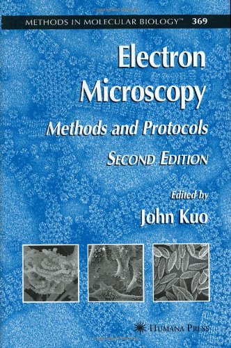
19 reviewsThe revised and expanded second edition of this book presents the newest technology in electron microscopy, while maintaining the practicality and accessibility of the acclaimed first edition. Like the first edition, this volume provides clear, concise instructions on processing biological specimens and includes discussion on the underlying principles of the majority of the processes presented. Electron Microscopy comprises two major areas of electron microscopy-transmission electron microscopy (TEM) and scanning electron microscopy (SEM). The TEM area covers several key techniques, including: conventional specimen preparation methods for cultured cells and biomedical and plant tissues; cryospecimen preparation by high-pressure freezing and cryoultramicrotomy negative staining and immunogold labeling techniques; and TEM crystallography and cryo-TEM tomography. The SEM area similarly attends to conventional-, variable pressure-, environmental-, and cryoscanning microscopy techniques, as well as the application of X-ray microanalysis. Protocols for the application of X-ray microanalysis to SEM and mass spectrometry conclude the volume.