Most ebook files are in PDF format, so you can easily read them using various software such as Foxit Reader or directly on the Google Chrome browser.
Some ebook files are released by publishers in other formats such as .awz, .mobi, .epub, .fb2, etc. You may need to install specific software to read these formats on mobile/PC, such as Calibre.
Please read the tutorial at this link. https://ebooknice.com/page/post?id=faq
We offer FREE conversion to the popular formats you request; however, this may take some time. Therefore, right after payment, please email us, and we will try to provide the service as quickly as possible.
For some exceptional file formats or broken links (if any), please refrain from opening any disputes. Instead, email us first, and we will try to assist within a maximum of 6 hours.
EbookNice Team
Status:
Available4.6
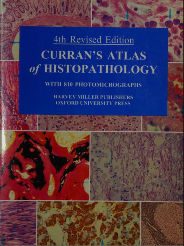
5 reviewsThis is the fourth edition of Professor Curran's well-known and widely used colour atlas of histopathology. The text has been completely revised and there have been additional immunohistological images added to the 804 full colour illustrations that make the atlas such a valuable reference
text for students and pathologists alike. The clarity of the text and the colour balance of the sharply-focused illustrations are unique. The contents of each illustration were carefully chosen and balanced for the structural importance. The boundaries of the pictures are defined with precision, to
make sure that very little space is wasted in the tissues. The general arrangement of the contents has been retained, with a chapter on each of the main systems or organs of the body. There is an introductory chapter of a general nature which demonstrates the more important reactions of the tissues
in disease and at the same time teaches the student the basic language of histopathology, thereby enabling him or her to read and assess the significance of changes in the tissue as revealed by microscopy. Most of the conditions are common or fairly common diseases, but occasional rare lesions are
included. This book is primarily an atlas, the primary purpose of which is to convey information in visual form. It is meant to complement existing textbooks. A new comprehensive index has been prepared for this edition. The book is intended primarily for undergraduate students but experience with
its predecessors suggests that it is likely to prove useful to postgraduate students in training in pathology or other clinical disciplines.
Contents
Preface
Acknowledgements
Methods
1. General Pathology
2. Alimentary Tract
3. Bones, Cartilage & Joints
4. Central Nervous System & Eye
5. Ear, Nose & Mouth
6. Endocrine Organs
7. Generative System, Female, including Breast
8. Generative System, Male
9. Heart & Arteries
10. Kidneys & Bladder
11. Liver, Gall Bladder & Pancreas
12. Lungs, Bronchi & Trachea
13. Lymphoid Tissue, Spleen & Blood
14. Skin
Index
4.6
31 reviews
4.6
12 reviews
5.0
14 reviews
5.0
7 reviews
4.4
25 reviews
4.5
17 reviews