Most ebook files are in PDF format, so you can easily read them using various software such as Foxit Reader or directly on the Google Chrome browser.
Some ebook files are released by publishers in other formats such as .awz, .mobi, .epub, .fb2, etc. You may need to install specific software to read these formats on mobile/PC, such as Calibre.
Please read the tutorial at this link. https://ebooknice.com/page/post?id=faq
We offer FREE conversion to the popular formats you request; however, this may take some time. Therefore, right after payment, please email us, and we will try to provide the service as quickly as possible.
For some exceptional file formats or broken links (if any), please refrain from opening any disputes. Instead, email us first, and we will try to assist within a maximum of 6 hours.
EbookNice Team
Status:
Available4.6
16 reviews
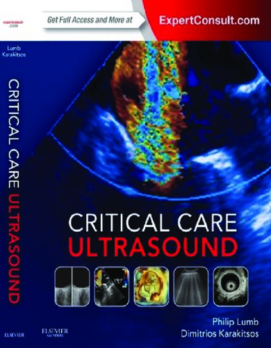
ISBN-10 : 1455753572
ISBN-13 : 9781455753574
Author: Philip D. Lumb
The increased use of ultrasound in critical care necessitates further training. Critical Care Ultrasound provides a straightforward, practical approach, an abundance of detailed ultrasound images provide step-by-step guidance on the principles and effective use of this important imaging modality in both diagnosis and assistance with specific procedures.
SECTION I: Fundamentals
Chapter 1: Fundamentals: Essential technology, concepts, and capability
Fundamentals: Principles, terms, and concepts
Equipment and imaging modes
Image quality and optimization
Artifacts
Ultrasound technique and safety issues
Scope and evolution of ultrasound imaging
Emergency ultrasound
Critical care ultrasound
The holistic approach ultrasound concept
Pearls and highlights
SECTION II: Neurocritical Care
Chapter 2: Transcranial doppler ultrasound in neurocritical care
Overview
Basic principles
Acoustic windows
Transcranial doppler interpretation
Transcranial doppler applications
Transcranial color-coded duplex (consultant level examination)
Pearls and highlights
Chapter 3: Transcranial doppler in aneurysmal subarachnoid hemorrhage: (CONSULTANT LEVEL EXAMINATION)
Overview
Vasospasm after aneurysmal subarachnoid hemorrhage
Transcranial doppler as a monitor for cerebral vasospasm
Indices and technical aspects of transcranial doppler ultrasonography
Interpretation of data from transcranial doppler
Limitations of transcranial doppler in the detection of vasospasm
Management strategy in aneurysmal subarachnoid hemorrhage
Transcranial doppler ultrasonography for traumatic vasospasm
Transcranial doppler as a marker of intracranial pressure and compliance
Pearls and highlights
Chapter 4: Transcranial doppler in the diagnosis of cerebral circulatory arrest: (CONSULTANT LEVEL EXAMINATION)
Overview
Transcranial doppler in the diagnosis of cerebral circulatory arrest
Pearls and highlights
Chapter 5: Use of transcranial doppler ultrasonography in the pediatric intensive care unit: (CONSULTANT LEVEL EXAMINATION)
Overview
Technique
Doppler measurements
Basic cerebrovascular hemodynamics
Transcranial color doppler imaging ultrasonography in the pediatric intensive care unit
Pearls and highlights
Chapter 6: Ocular ultrasound in the intensive care unit: (CONSULTANT LEVEL EXAMINATION)
Overview
Equipment and settings
General scanning technique and primary views
Trauma
Optic nerve sheath diameter measurements and new quality criteria
Pupillary light reflex assessment
Additional specialist evaluation
Safety aspects
Pearls and highlights
Chapter 7: General chest ultrasound in neurocritical care
Overview
Cardiovascular evaluation in neurocritical care
Pulmonary evaluation: Neurogenic pulmonary edema
Pearls and highlights
SECTION III: Vascular Ultrasound
Chapter 8: Overview of the arterial system
The arterial system
Disorders
Pearls and highlights
Chapter 9: Ultrasonography for deep venous thrombosis
Background
Anatomy
Basic ultrasound techniques
Diagnostic criteria
Ultrasound examination strategies in the intensive care unit
Pearls and highlights
Chapter 10: Ultrasound-guided central venous access: The basics
Overview
Advantages of ultrasound-guided vascular access
The ultrasound transducer
The display
Image optimization
Vascular scanning
Needle orientation
Selection of the site of cannulation
Ultrasound-guided vascular access techniques and current recommendations
Pearls and highlights
Chapter 11: Ultrasound-guided vascular access: Trends and perspectives
Patient and technical considerations
Preprocedural tips
Variations in technique
Intraprocedural and postprocedural tips
Simulation training and echogenic technology
Pearls and highlights
Chapter 12: How to choose the most appropriate ultrasound-guided approach for central line insertion: Introducing the rapid central venous assessment protocol
Overview
The rapid central vein assessment
Criteria for choosing the appropriate vein according to the rapid central vein vascular assessment protocol
Pearls and highlights
Chapter 13: Pediatric ultrasound-guided vascular access
General considerations and ultrasound evaluation of peripheral and central veins in pediatric patients (preprocedural scanning)
Instrumentation and kits used for ultrasound-guided catheterization in neonates and children
Technique of ultrasound-guided central venous catheterization in neonates and small infants (younger than 3 months)
Technique of ultrasound-guided central venous catheterization in infants and small children (3 months to 6 years old)
Technique of ultrasound-guided insertion of peripheral lines in pediatric patients
Peripherally inserted central venous catheters in children
Pearls and highlights
Chapter 14: Ultrasound-guided peripheral intravenous access
Pearls and pitfalls
Chapter 15: Ultrasound-guided placement of peripherally inserted central venous catheters
Overview
Ultrasound anatomy of the arm veins
The safe insertion protocol for peripherally inserted central catheters
Pearls and highlights
Chapter 16: Ultrasound-guided arterial catheterization
Overview
Procedure and instrumentation
Site-specific tips
Pearls and highlights
Chapter 17: Intravascular ultrasound: (CONSULTANT-LEVEL EXAMINATION)
Overview
Clinical applications of intravascular ultrasound for percutaneous coronary interventions
Intravascular ultrasound for carotid revascularization
Pearls and highlights
Chapter 18: Ultrasound-guided placement of inferior vena cava filters: (CONSULTANT-LEVEL EXAMINATION)
Technique of bedside intravascular ultrasound–guided inferior vena cava filter placement
Pearls and highlights
SECTION IV: General Chest Ultrasound
Chapter 19: Lung ultrasound: The basics
Overview
The normal lung
Evaluation for pneumothorax
Evaluation for pulmonary edema (Interstitial Syndrome)
Evaluation for consolidation (alveolar syndrome)
Evaluation for pleural effusion
Goal-directed applications
Limitations
Pearls and highlights
Chapter 20: Pleural ultrasound
Overview
Identification of anatomic structures
Lung sliding
Lung pulse
Pneumothorax
Pleural effusions
Solid pleural abnormalities
Pleuroparenchymal disorders
Diagnostic and interventional applications of pleural ultrasound
Pitfalls and limitations
Pearls and highlights
Chapter 21: Lung ultrasound in trauma
Overview
The technique
The normal pattern
Pneumothorax
Lung contusion
Hemothorax
Pearls and highlights
Chapter 22: Lung ultrasound in acute respiratory distress syndrome (ARDS)
Overview
Lung ultrasound b-lines and extravascular lung water
Ultrasonographic signs of acute respiratory distress syndrome
Acute respiratory distress syndrome versus acute cardiogenic pulmonary edema: Differential diagnosis by lung ultrasound
The integration of lung ultrasound in acute respiratory distress syndrome management
Technical issues
Pearls and highlights
Chapter 23: Lung ultrasound in mechanically ventilated patients
Overview
Recruitment/positive end-expiratory pressure (PEEP)
Screening for complications of mechanical ventilation
The difficult-to-wean patient
Lung ultrasound with alternative forms of ventilation
Pearls and highlights
isccm manual of critical care ultrasound
manual of emergency and critical care ultrasound
fellowship in critical care ultrasound
comprehensive critical care ultrasound
critical care ultrasound course 2025
Tags: Critical Care, Ultrasound, Philip Lumb, further training
5.0
8 reviews
4.4
22 reviews
5.0
11 reviews
5.0
13 reviews
4.7
13 reviews
4.4
5 reviews

4.4
40 reviews