logo
Product categories

EbookNice.com

Most ebook files are in PDF format, so you can easily read them using various software such as Foxit Reader or directly on the Google Chrome browser.
Some ebook files are released by publishers in other formats such as .awz, .mobi, .epub, .fb2, etc. You may need to install specific software to read these formats on mobile/PC, such as Calibre.

Please read the tutorial at this link.  https://ebooknice.com/page/post?id=faq


We offer FREE conversion to the popular formats you request; however, this may take some time. Therefore, right after payment, please email us, and we will try to provide the service as quickly as possible.


For some exceptional file formats or broken links (if any), please refrain from opening any disputes. Instead, email us first, and we will try to assist within a maximum of 6 hours.

EbookNice Team
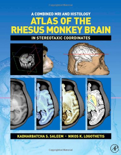
(Ebook) A Combined MRI and Histology Atlas of the Rhesus Monkey Brain in Stereotaxic Coordinates by Kadharbatcha S. Saleem, Nikos K. Logothetis ISBN 9780080467771, 9780123725592, 0080467776, 0123725593

  • SKU: EBN-1915656
Zoomable Image
$ 32 $ 40 (-20%)

Status:

Available

4.3

20 reviews
Instant download (eBook) A Combined MRI and Histology Atlas of the Rhesus Monkey Brain in Stereotaxic Coordinates after payment.
Authors:Kadharbatcha S. Saleem, Nikos K. Logothetis
Pages:336 pages.
Year:2006
Editon:1st
Language:english
File Size:33.67 MB
Format:pdf
ISBNS:9780080467771, 9780123725592, 0080467776, 0123725593
Categories: Ebooks

Product desciption

(Ebook) A Combined MRI and Histology Atlas of the Rhesus Monkey Brain in Stereotaxic Coordinates by Kadharbatcha S. Saleem, Nikos K. Logothetis ISBN 9780080467771, 9780123725592, 0080467776, 0123725593

This atlas maps the detailed architectonic subdivisions of the cortical and subcortical areas in the macaque monkey brain using high-resolution magnetic resonance (MR) images and the corresponding histology sections in the same animal. This book presents the detailed mapping of the architectonic areas in the horizontal plane of sections with reference to the MRI that has not been reported previously in macaque monkeys. In the second part of the atlas, the coronal plane is presented using the same technique. A third part shows the quick identification of several important cortical and subcortical areas (around 30 areas) in horizontal, coronal and sagittal MR images. This atlas is unlike anything else available as it includes and compares each section to imaging data. This is a significant progress, as the vast majority of research in the field now routinely work with fMRI images. ?· Provides the first combined MRI and Histology maps of the cortical and subcortical areas of any non-human primate species?· Shows the first detailed delineations of the cortical and subcortical areas in both horizontal and coronal planes in the same animal using five different staining methods?· Illustrates the entire dorsoventral extent of the left hemisphere in 47 horizontal MRI and photomicrographs matched with 47 detailed diagrams (Chapter 3) ?· Presents the full rostrocaudal extent of the right hemisphere in 76 coronal MRI and photomicrographs, and 76 corresponding drawings (Chapter 4)?· Illustrates the selected cortical and subcortical areas in horizontal, coronal and sagittal MRI planes (Chapter 5)?· Provides the sterotaxic grid derived from the in-vivo MR image?· Likely to become a standard reference for anatomical, physiological, and functional imaging studies in primates (fMRI, PET and MEG)
*Free conversion of into popular formats such as PDF, DOCX, DOC, AZW, EPUB, and MOBI after payment.

Related Products